Home
/ Figure 1 Animal Cell - 1 The Structure Labeled A Are Found In Bothtypes Of Cells The Name Of The Structure Is2 Unlike Brainly Ph - (a)€€€€€calculate the magnification of the microscope.
Figure 1 Animal Cell - 1 The Structure Labeled A Are Found In Bothtypes Of Cells The Name Of The Structure Is2 Unlike Brainly Ph - (a)€€€€€calculate the magnification of the microscope.
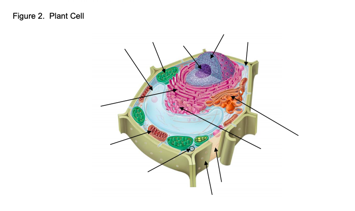
Figure 1 Animal Cell - 1 The Structure Labeled A Are Found In Bothtypes Of Cells The Name Of The Structure Is2 Unlike Brainly Ph - (a)€€€€€calculate the magnification of the microscope.. Choose the answers from the box. What does this mean about the cells that are found in each of these living things? (a) a plasmodesma is a channel between the cell walls of two adjacent plant cells. Cells and tissues that comprise the stomach. The plant cell has a cell wall, chloroplasts, plastids, and a central vacuole—structures not found in animal cells.
Transport vesicles hab e rdar o lo que 22. 1 what is structure a? All cells are surrounded by a. Based on the structure of the plasma membrane, it is regarded as the fluid mosaic model. Figure 1 shows two cells, x and y.
Figure 1 From Molecular Control Of Animal Cell Cytokinesis Semantic Scholar from d3i71xaburhd42.cloudfront.net This problem has been solved! Color and /abe/the microtubules are shaped like soda straws and give the nucleus and cell its shape. Figure 1 shows an animal cell viewed using a microscope figure 1 the cell contains a nucleus. The animal cell contains a. 3 figure 2 shows a sperm. cell membrane cell wall chloroplast cytoplasm nucleus only the animal cell contains a _____. (a)€€€€€calculate the magnification of the microscope. Organelles of the animal cell and their function.
What does this mean about the cells that are found in each of these living things?
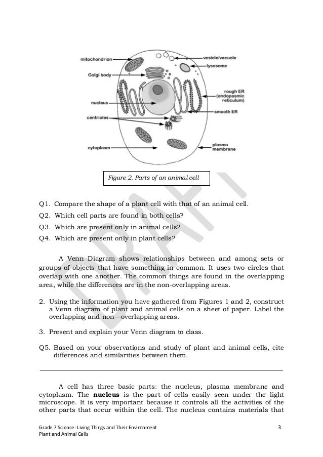
Animal cells differ from plant cells in several regards though, including the lack of vacuoles, chloroplasts, and cell walls. These figures show the major organelles and other cell components of (a) a typical animal cell and (b) a typical eukaryotic plant cell. Figure 1 summary of events during cytokinesis. This problem has been solved! Based on the structure of the plasma membrane, it is regarded as the fluid mosaic model. The diagram does not show a cell wall layer and a centrally located vacuole is absent as well, which is the characteristics of a plant cell. Figure 1 shows an animal cell viewed using a microscope figure 1 the cell contains a nucleus. The centriole is the dense center of the centrosome. Choose the answers from the box. (a generalised cell shows all the structures that are typically found in a cell.) figure 1.6 shows some actual human cells and figure 1.7 shows an actual plant cell Start studying apologia module 6, figure 6.1\\animal cell. 2 what is structure b? In an animal cell, the cell membrane functions by providing shape and protects the inner contents of the cell.
Learn vocabulary, terms, and more with flashcards, games, and other study tools. What does this mean about the cells that are found in each of these living things? 1 mark tick one box. This is in stark contrast to the neuron in the human body, which is just 100 microns across. (d) gap junctions act as channels between animal cells.
Gr7module2forstudents 120920041248 Phpapp01 1 from image.slidesharecdn.com Made first observation of cells (cork) c) theodor schwann (1830‟s): Start studying apologia module 6, figure 6.1\\animal cell. What does this mean about the cells that are found in each of these living things? List the colors you actually used in your drawings if they are different from the ones shown. Figure 1.1 (a) a simple animal cell and (b) a plant leaf cell 58750_p012_053.indd 14 5/7/16 12:15 pm google search: This is in stark contrast to the neuron in the human body, which is just 100 microns across. The largest known animal cell is the ostrich egg, which can stretch over 5.1 inches across and weighs about 1.4 kilograms. 1 mark tick one box.
Animal cell diagram vacuole :
(a) a plasmodesma is a channel between the cell walls of two adjacent plant cells. (b) tight junctions join adjacent animal cells. These figures show the major organelles and other cell components of (a) a typical animal cell and (b) a typical eukaryotic plant cell. 1 what is structure a? 0 1 figure 1 shows an animal cell. Plant cells do not have lysosomes or centrosomes. Choose the answers from the box. Image from purves et al., life: (a generalised cell shows all the structures that are typically found in a cell.) figure 1.6 shows some actual human cells and figure 1.7 shows an actual plant cell Figure 1 shows a cell viewed through a light microscope. Comments (0) answer & explanation. The science of biology, 4th edition, by sinauer associates (www.sinauer.com) and wh freeman (www.whfreeman.com), used with permission. Transport vesicles hab e rdar o lo que 22.
Only animal cells have centrosomes. Image from purves et al., life: See animal cell stock video clips. (a) a plasmodesma is a channel between the cell walls of two adjacent plant cells. Animal cell diagram vacuole :
Cell Structures And Functions Use Www Cellsalive Com Chegg Com from media.cheggcdn.com Figure 1 € the size of the real cell is 0.03 mm. Expert answer 100% (1 rating) previous question next question transcribed image text from this question. 0 1 figure 1 shows an animal cell. By knowing what organelles animal cells have and their general shapes, you can easily draw an animal cell. The plant cell has a cell wall, chloroplasts, plastids, and a central vacuole—structures not found in animal cells. Label the microtubules inside the nucleus. To check if you have understood the cell parts, draw a blank animal cell diagram and try to fill in the different parts without referring to the labeled one given here. 1 mark tick one box.
Figure 1 € the size of the real cell is 0.03 mm.
Below you can find a list will all of them (animal cell organelles and their functions) with and image/diagram to help you visualize where they are and how they look within the cell. Only animal cells have centrosomes. This problem has been solved! 3 specimen material 0 1. 2 what is structure b? The largest known animal cell is the ostrich egg, which can stretch over 5.1 inches across and weighs about 1.4 kilograms. The diagram represents the animal cell as it shows a centrally located nucleus and cell membrane as its outer boundary. Figure 1 shows a cell viewed through a light microscope. cell membrane cell wall chloroplast cytoplasm nucleus only the animal cell contains a _____. All cells are surrounded by a. (a)€€€€€calculate the magnification of the microscope. In an animal cell, the cell membrane functions by providing shape and protects the inner contents of the cell. First observed of animal cells • lack of cell wall delayed discovery (made viewing difficult…) 1) every living organism is made up of 1 or more cells
1 mark tick one box animal cell figure. Chromosome and dna anatomy of animal cell animal cell diagram animal cell anatomy animal cells diagram dna and cells dna in cell dna cell chromosome plant cell animal cell animal cell structure.
Share :
Post a Comment
for "Figure 1 Animal Cell - 1 The Structure Labeled A Are Found In Bothtypes Of Cells The Name Of The Structure Is2 Unlike Brainly Ph - (a)€€€€€calculate the magnification of the microscope."


Post a Comment for "Figure 1 Animal Cell - 1 The Structure Labeled A Are Found In Bothtypes Of Cells The Name Of The Structure Is2 Unlike Brainly Ph - (a)€€€€€calculate the magnification of the microscope."